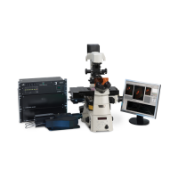
A1 MP Multiphoton Confocal
Send a free RFQ and get the supplier's best price.
Trade with confidence on Abraa
Abraa güvenli ticaret sistemlerini kullanarak güvenli ve emniyetli ödemeler
Supplier verification not completed
Pay only through Abraa to stay protected. Never pay a supplier by direct transfer.
Secure Payment
Pay through Abraa, never by direct transfer to a supplier.
Product Inspection
Book a check before shipping to confirm quality and quantity.
Alıcı Koruması
Get support and refunds if goods arrive not as described.
Verified Suppliers
Licence, address and on-site checks by the Abraa team.
Ürün Özellikleri
| JLM Medical Instruments LLC | |
| New | |
| YES |
Product Descriptions
Streamlined multiphoton confocal for deeper, faster multiphoton imaging.
Nikon’s A1 MP+ multiphoton confocal microscope is a unique multiphoton imaging system featuring a high resolution galvanometer scanner. New 4 channel non-descanned detectors with higher sensitivity, reduced dark current and broad spectral range allow for real time unmixing of closely spaced probes for accurate and high-contrast spectral imaging. This is especially important in multiphoton microscopy because of the overlap of emission spectra of probes and autofluorescence, which is often unavoidable when using a single laser line.
Deep Specimen Imaging with High-sensitivity Non-Descanned Detectors (NDD)
The fluorescence emissions from deep within a specimen are highly scattered in multiphoton excitation, and therefore the conventional detector using a pinhole cannot provide bright fluorescent images. The episcopic NDD in the A1 MP+ is located close to the back aperture of the objective to detect the maximum amount of scattered emission signals from deep within living specimens.
In vivo image of deep areas of cerebral cortex of a mouse
The cerebral cortex of an H-line 5-week-old mouse was studied with the open skull method. The entire shape of dendrites of pyramidal cells in layer V expressing EYFP were visualized from the bottom layer into a superficial layer. In addition, the fluorescence signal of white matter in deeper areas was also studied.
| Left | 3D reconstruction image |
Photographed with the cooperation of: Dr. Tomomi Nemoto, Dr. Shigenori Nonaka, Dr. Takeshi Imamura, |
|
| Right | Z-stack images | ||
| Top: dendrites located in superficial layers in the layer V pyramidal cells 25 µm from the surface |
|||
| Middle: basal dendrites in the layer V pyramidal cells 625 µm from the surface |
|||
| Bottom: fluorescence from white matter Excitation wavelength: 950 nm Objective: CFI75 Apo 25xW MP (NA 1.10 WD 2.0) |
3D volume rendering images
1300nm Imaging
Deep brain imaging in in vivo mouse with the GaAsP NDD compatible with the 1300 nm wavelength
Mouse brain in vivo dual color imaging with the GaAsP NDD compatible with the 1300 nm wavelength
Nikon High-NA Objectives are Ideal for Multiphoton Imaging
Auto Laser Alignment when Changing Multiphoton Excitation Wavelength
Increased flexibility and ease of use
for more information please visit :
https://www.nikoninstruments.com/Products/Multiphoton/A1MP-Multiphoton
 \