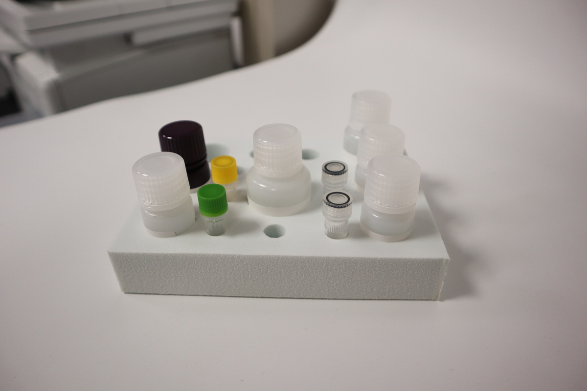
Human GLP1(Glucagon-like peptide 1) ELISA Kit
التوفر: في المستودع
Trade with confidence on Abraa
طريقة دفعة آمنة وموثوقة باستخدام منصة عبرة للدفع عبر الانترنت
Supplier verification not completed
Pay only through Abraa to stay protected. Never pay a supplier by direct transfer.
Secure Payment
Pay through Abraa, never by direct transfer to a supplier.
Product Inspection
Book a check before shipping to confirm quality and quantity.
حماية المشتري
Get support and refunds if goods arrive not as described.
Verified Suppliers
Licence, address and on-site checks by the Abraa team.
مواصفات المنتج
| الماركة | Feiyuebio |
مواصفات المنتج
Reactivity:Human
Assay range:12.35–1,000pg/mL
Sensitivity:< 4.92pg/mL
Detection Method:Competitive Inhibition
Storage:2–8 ℃ for 6 months
CV (%):Intra-Assay: CV<8%; Inter-Assay: CV<10%
Note:For Research Use Only
GLP1(Glucagon-like peptide 1) Basic information
GLP-1 is a 30- or 31-amino-acid-long peptide hormone deriving from the tissue-specific posttranslational processing of the proglucagon peptide. It is produced and secreted by intestinal enteroendocrine L-cells and certain neurons within the nucleus of the solitary tract in the brainstem upon food consumption. The initial product GLP-1 (1–37) is susceptible to amidation and proteolytic cleavage, which gives rise to the two truncated and equipotent biologically active forms, GLP-1 (7–36) amide and GLP-1 (7–37). Active GLP-1 protein secondary structure includes two α-helices from amino acid position 13–20 and 24–35 separated by a linker region.
Human GLP1(Glucagon-like peptide 1) ELISA Kit ELISA Kit ELISA Kit test method
This kit was based on Competitive-ELISA detection method. The microtiter plate provided in this kit has been pre-coated with target. During the reaction, target in the sample or standard competes with a fixed amount of target on the solid phase supporter for sites on the Biotinylated Detection Antibody specific to target. Excess conjugate and unbound sample or standard are washed from the plate, and HRP-Streptavidin (SABC) is added to each microplate well and incubated. Then TMB substrate solution is added to each well. The enzyme-substrate reaction is terminated by the addition of a sulphuric acid solution and the color change is measured spectrophotometrically at a wavelength of 450nm. The concentration of target in the samples is then determined by comparing the OD of the samples to the standard curve.
 \
منتجات مشابهة
View all


